Ocular surface analysis and automatic non-invasive assessment of tear film breakup location, extension and progression in patients with glaucoma. BMC Ophthalmol. 2020 Jan 6;20(1):12. doi: 10.1186/s12886-019-1279-7. PMID: 31906897; PMCID: PMC6945571.
Summary
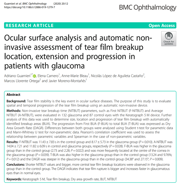
Tear film stability is the key element in ocular surface diseases. The aim of this study is to assess the spatial and temporal progression of tear film breakdown using an automated non-invasive device.
Method: Non-invasive tear film breakage (TFTB) measurements, such as first TFTB (F-TFTB) and average TFTB (A-TFTB), were performed on 132 glaucomatous eyes (glaucoma group) and 87 healthy eyes (control group) with the Keratograph 5M device. Further analysis of these data allowed the size, location and progression of tear film breakage with automatically identified breakage zones (BUA) to be determined. The progression from the first BUA (F-BUA) to the total BUA (T-BUA) was expressed as the dry area growth rate (DAGR).
Results: F-NITBUT was 11.43 ± 7.83 seconds in the control group and 8.17 ± 5.73 seconds in the glaucoma group (P = 0.010). A-NITBUT was 14.04 ± 7.21 seconds in the control group and 11.82 ± 6.09 seconds in the glaucoma group, respectively (P = 0.028). F-BUA was higher in the glaucoma group than in the control group (2.73 and 2.28; P = 0.022) and was more frequently located in the centre of the cornea in the glaucoma group (P = 0.039). T-BUA was also higher in the glaucoma group than in the control group (13.24 and 9.76%; P = 0.012) and DAGR was more pronounced in the glaucoma group than in the control group (34.38° and 27.15°; P = 0.009).
Conclusions: Shorter NITBUT values and larger and more central tear film breakage points were observed in the glaucoma group compared to the control group. The DAGR indicates that tear film breakage is greater and increases more rapidly in glaucomatous eyes than in normal eyes. This implies that...
